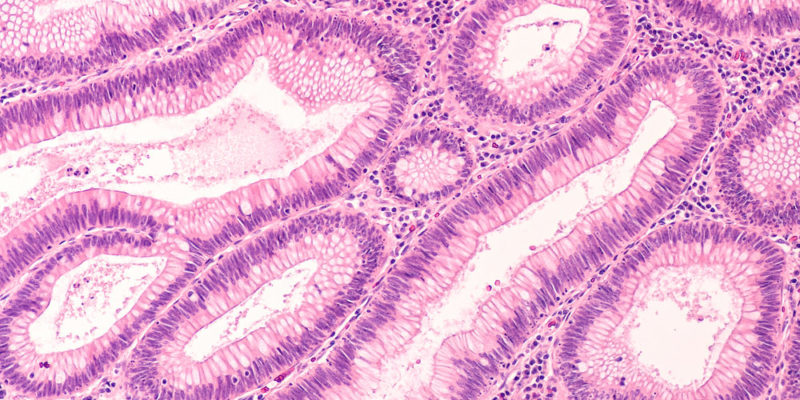
Histology is the study of the microscopic organization of tissues. For medical students, it is not just about “seeing cells under the microscope,” but about learning to recognize patterns: where there is a lining, where there is support, where there is contraction, and where there is signal conduction. That visual vocabulary shows up again later in pathology, physiology, anatomy, and clinical medicine.126
Histology is not merely a “basic science” subject. It is the point where anatomy, physiology, and pathology begin to speak the same language: the language of tissue organization.12 Anatomy shows where organs are and how they relate in space; histology shows how those organs are built from the inside and why that construction supports normal function.126
That difference may seem theoretical at first, but it quickly becomes practical. Understanding epithelium, stroma, muscle, and nervous tissue helps explain why a mucosa absorbs, why an organ becomes fibrotic, why a gland secretes, and why some lesions impair function even before they cause major macroscopic change.12
In medical practice, histology is also a direct foundation of diagnosis. Biopsies, surgical specimens, and much of pathology depend on recognizing normal architecture and structural alteration. Before asking “what is the disease?”, it is often more accurate to ask which tissue is altered and how it deviates from the expected pattern.25
When a slide is placed under the microscope, the goal is not to identify every isolated cell, but to understand the architecture of the whole sample. Histology asks how cells are organized, what their relationship is to the extracellular matrix, and whether there is a free surface, a lumen, a basement membrane, fibers, vessels, or clearly defined layers.12
That is why the classification into four basic tissues remains so useful: it organizes the reading process. Epithelial tissue dominates surfaces and glands; connective tissue supports, connects, and fills; muscle produces force; and nervous tissue integrates and conducts information.16
In other words, histology is less a list of names and more an exercise in recognizing spatial relationships. What you see under the microscope is function translated into form.12
One of the biggest difficulties at the beginning is trying to identify the diagnosis or the organ before understanding the basic organization of the specimen. In general, it works better to follow a short and repeatable mental sequence.25
| Guiding question | What to look for first | What it usually suggests |
|---|---|---|
| Is there a surface or a lumen? | Continuous lining, apical border, glands | Epithelium |
| Is there a lot of extracellular space? | Fibers, ground substance, vessels | Connective tissue |
| Are the cells elongated and arranged in bundles? | Striations, peripheral or central nuclei | Muscle |
| Are there large cell bodies or wavy bundles? | Neurons, glia, peripheral nerves | Nervous tissue |
Starting at the lowest magnification also helps. At that level, you can recognize whether the section has layers, whether there is mucosa, capsule, bundles, septa, or transition zones. Only then is it worth increasing magnification to inspect nuclear details, cytoplasm, fibers, and cell borders.25
Another useful strategy is to mentally separate what is more cellular from what is more matrix-rich. Epithelia and many muscle tissues tend to be more compact; connective tissues, by definition, show more extracellular matrix; nerves combine specialized cells with a glial support background.148
The appearance of a slide depends not only on the tissue itself, but also on how it was processed. In routine histology, the material goes through fixation, processing, paraffin embedding, microtome sectioning, and staining.5
Fixation preserves structure and reduces autolysis and degradation. After that, the tissue is dehydrated, embedded in paraffin, and cut into thin sections so that light can pass through it. Only then does the slide receive stain, and the most important one in day-to-day practice is Hematoxylin and Eosin (H&E).5
In H&E, hematoxylin marks more basophilic structures, especially nuclei, in blue to purple tones; eosin imparts pink tones to cytoplasm, collagen, and several matrix proteins.5 That simple contrast is already enough to answer many of the first questions in histology: where the cells are, how they are distributed, and how they relate to the stroma.
It is worth remembering that not everything on the slide is “pure biology.” Folds, retraction, artificial spaces, and small tears may be processing artifacts. Learning histology also means learning to question what looks too strange to be normal structure.25
Hematoxylin and Eosin remains the best entry point because it gives an overall view of the tissue.5 But in practice, not every question is answered by a general stain. When the goal is to highlight a specific component more clearly, so-called special stains become useful.5
| Stain | What it highlights | When it usually helps |
|---|---|---|
| H&E | Overall view of nucleus, cytoplasm, and matrix | First reading of most slides |
| PAS | Carbohydrate-rich structures and basement membranes | Mucin, brush border, renal tubules, and some matrix details |
| Masson's trichrome | Contrast between muscle and collagen | Fibrosis, scar, and stromal organization |
| Silver-based methods | Certain fibers and special structures | Situations in which H&E does not separate the detail of interest well |
At the beginning, it is not worth memorizing a catalog of techniques. What matters most is understanding the logic: H&E answers the general question; special stains are used when a more specific question needs a clearer answer.5
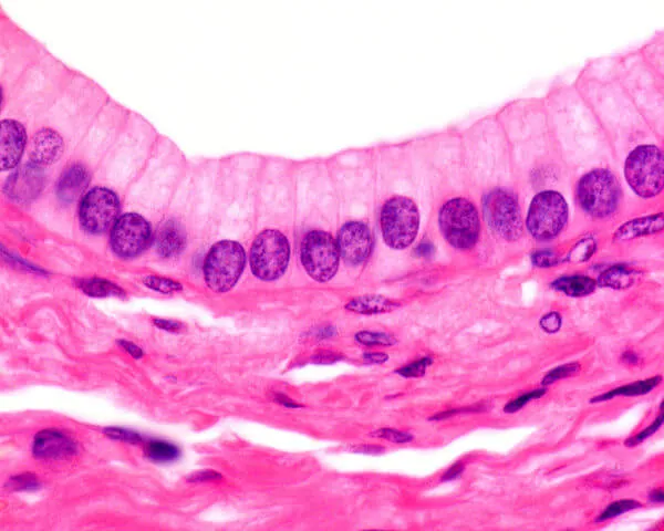
Epithelial tissue lines external surfaces and internal cavities, and it also forms glands. Its cells are tightly packed, with little extracellular matrix, and they rest on a basement membrane that separates the epithelium from the underlying connective tissue.13
Functionally, epithelium can protect, absorb, secrete, and transport substances. Apical specializations help with interpretation: microvilli favor absorption; cilia participate in moving material along the surface; and cell shape itself often reflects the dominant task.3
Classically, epithelium is described by the number of layers and the shape of the superficial cells. That system is simple, but it remains extremely useful under the microscope.13
| Criterion | Main types | Classic examples |
|---|---|---|
| Number of layers | Simple, stratified, pseudostratified, transitional | Alveoli, epidermis, respiratory tract, bladder |
| Cell shape | Squamous, cuboidal, columnar | Endothelium, renal tubules, intestine |
Connective tissue does almost the opposite of epithelium: it is defined less by cell apposition and more by the richness of the extracellular matrix. That matrix includes ground substance and fibers, especially collagen, reticular, and elastic fibers.14
Its function goes far beyond “filling space.” Connective tissue supports organs, participates in immune defense, allows exchange with vessels, stores energy when specialized as adipose tissue, and serves as the basis for cartilage, bone, and blood.4
On a slide, a good shortcut is to look for the stroma: lighter or more fibrous regions between epithelia, muscle bundles, or glandular structures. When that stroma is loose, there is more space and a greater variety of cells; when it is dense, fibers predominate and determine the tissue's mechanical resistance.124
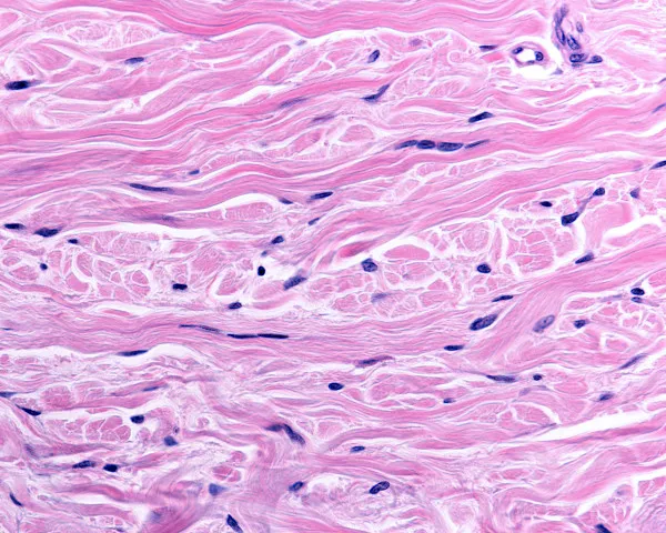
Muscle tissue is specialized for contraction. Although all of its types share that function, they are clearly distinguishable in conventional microscopy.17
Skeletal muscle is made of long, striated, multinucleated fibers, usually with peripheral nuclei. Cardiac muscle is also striated, but it has branched cells, central nuclei, and intercalated discs, which help synchronize electrical activity. Smooth muscle has no visible striations in routine light microscopy; its cells are spindle-shaped, with elongated central nuclei, and it appears in the walls of viscera and vessels.27
| Muscle type | Appearance on H&E | Where it usually appears |
|---|---|---|
| Skeletal | Obvious striations, long fibers, peripheral nuclei | Voluntary muscles |
| Cardiac | Striated, branched cells, central nuclei | Myocardium |
| Smooth | No striations, spindle-shaped cells | Intestine, vessels, uterus, airways |
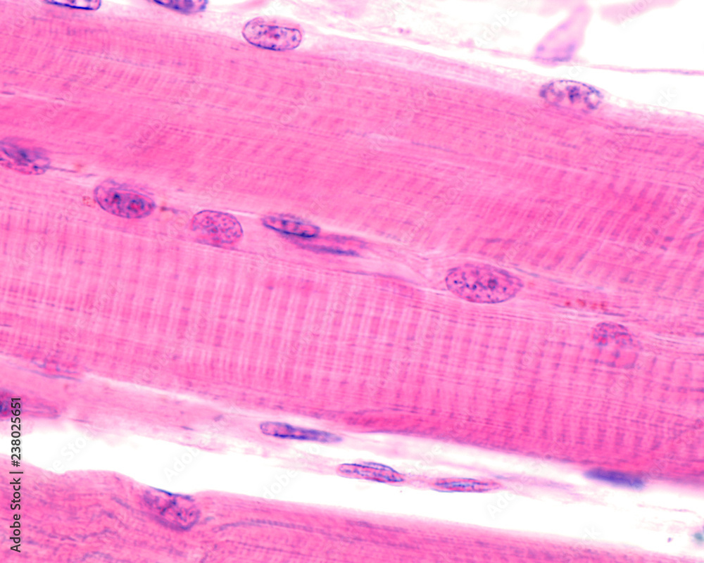
Nervous tissue is composed of neurons and support cells, the glia. In histologic practice, this means recognizing both cells specialized for signal conduction and a microenvironment that supports metabolism, myelin, homeostasis, and responses to injury.28
Neurons have a cell body, dendrites, and an axon; glia include, among other types, astrocytes, oligodendrocytes, Schwann cells, microglia, and ependymal cells.8 Not all of these elements are equally easy to distinguish on H&E, but the overall logic matters: nervous tissue is rarely just “the neuron.” It is always neuron plus support.
In peripheral nerves, it is common to see wavy bundles accompanied by connective tissue sheaths. In the central nervous system, the reading changes: gray matter, white matter, neuronal cell bodies, glia, and myelin each enter the picture with their own patterns.28
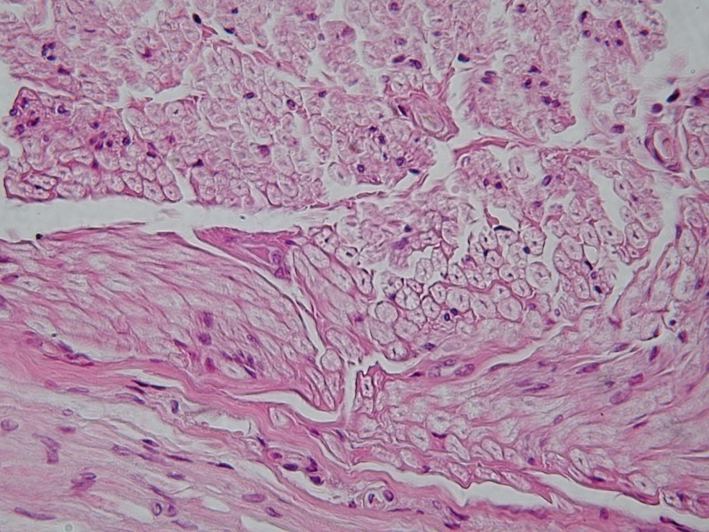
A real organ almost never shows only one tissue type. The value of histology grows precisely when you begin to see how those tissues combine to produce function.126
The intestine is a good example. Facing the lumen, there is simple columnar epithelium with absorptive and secretory functions; just beneath it lies the lamina propria, a loose connective tissue rich in cells and vessels; deeper in the wall there is smooth muscle organized into layers; and between those layers there are elements of the enteric nervous system that coordinate motility and secretion.1278
That integrated reading changes the way you study. Instead of memorizing “epithelial tissue,” “connective tissue,” and “muscle tissue” as separate boxes, you begin to understand that an organ is a recurring composition of those basic blocks arranged in specific patterns.6
| Tissue | What to look for first | Dominant function |
|---|---|---|
| Epithelial | Continuous lining, tightly packed cells, basement membrane | Protect, absorb, secrete |
| Connective | Extracellular matrix, fibers, vessels, more widely spaced cells | Support, connect, nourish, defend |
| Muscle | Elongated cellular bundles, with or without striations | Contract and generate force |
| Nervous | Neurons, glia, nerve bundles, myelin | Integrate and conduct information |
If histology feels abstract at first, it helps to simplify the question: which tissue predominates here, and why is it organized this way? That shift in approach is often what turns the slide into reasoning.
Clinical correlation
In diagnostic practice, the first question in front of a biopsy is almost never “what is the disease?” but rather “which compartment is altered?”. Recognizing epithelium, connective stroma, muscle, and nervous tissue reduces error and organizes slide reading even before any immunohistochemistry is considered.238
Turn this content into flashcards, mock exams, and AI-guided review. Start free on the Básico plan, no card required. The platform is available in Portuguese.
Epithelium usually forms continuous linings or glands, with tightly packed cells and little extracellular matrix. Connective tissue, by contrast, has more extracellular space, fibers, and more dispersed cells.
Anatomy describes the body at a macroscopic scale; histology deepens that study at a microscopic scale, showing how cells, extracellular matrix, and layers are organized inside tissues and organs.12
At minimum, it is useful to understand the logic of PAS, Masson's trichrome, and silver-based methods. They become useful when the question is not only “what is the general architecture?” but “which component do I need to highlight better?”.5
Content based on peer-reviewed academic literature and internationally recognized reference sources.